 |
|
|
|
 |
|
|
|
|
|
|
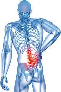
| Low Back Pain: Posture and Movement Analysis |
작성자 : 안성현 작성일 : 2015-03-24 |
|
|
|
|
|
Low Back Pain: Posture and Movement AnalysisBy Jeffrey Tucker, DC, DACRB Editor's note: This is a follow-up to Dr. Tucker's Jan. 15 article on the static postural pelvic exam.
When performing static and dynamic movement analysis of the lumbopelvic hip area, begin with standing visual posture analysis of the pelvis, and then perform lumbar range of motion and assess what you might see during normal versus abnormal lumbar flexion motion. Continue to the supine position and use common orthopedic tests to help determine muscle function that guides your diagnosis and corrective exercise strategy.
Visual Analysis (Standing) - Normal standing position: a forward convex curve (lordosis) of the lumbar spine of 25-35 degrees.
- Lateral tilt is observed if the iliac crest height is different (typically more than ½ inch). This may indicate a leg discrepancy, lumbar-region pathology, SI joint pathology, or shortness of the quadratus lumborum or latissimus dorsi muscles.
- Lateral shift is observed if the pelvis is shifted laterally to trunk. This may indicate lumbar-region pathology, short hip adductors (on the side of the shift) or weakened hip abductors (on the contralateral side).
- Rotation is observed if one ASIS is anterior of the contralateral ASIS; for example, the left innominate is more anteriorly tilted and forwardly rotated, with the right more posteriorly tilted and backwardly rotated. This position puts the right hip into internal rotation, adduction and extension; and the left hip compensatorily into external rotation, abduction, and flexion. This may contribute to lumbar-region pathology, SI joint pathology or an overactive TFL / ITB.
- Anterior pelvic tilt is observed with increased lumbar lordosis. This may indicate hip flexor (psoas, rectus femoris) and lumbar extensor hypertonicity or shortness; or imbalances in muscle strength and length, such as underactive glutes / hamstrings / abdominals. If there is excessive visceral fat, especially around the abdomen, this can lead to a lordotic posture. Anterior pelvis tilt is a common imbalance that leads to low back pain (facet syndrome), especially when the abdominals, which help support the spine, are weakened.
- Posterior pelvic tilt is observed in a flat back or decreased lumbar lordosis. This may indicate overactive / tight hamstrings / gluteal muscles. When the patient has a flat lumbar spine, the flexibility of the hips becomes particularly important. During forward bending, the individual with a flat lumbar spine must immediately flex the hips to avoid excessive flexion of the spine. If this can't be achieved, a low back pain syndrome may develop from excessive and repetitive lumbar flexion.
 Toe Touch (Standing) Toe Touch (Standing)
Next, ask the patient to perform lumbar range of motion. We are particularly interested in lumbar flexion movement. Ask the patient to "Bend over and touch your toes." The following represent some of the toe-touch presentations: - Normal lumbar flexion: The initial movement is the posterior sway of the pelvis, which allows the center of gravity to remain within the base of support. As the hips start to flex, the lumbar spine begins to reverse its inward curve, with further hip flexion completing the movement. The motion of lumbar flexion is approximately 60-70 degrees. Remember that the lumbar spine is in 30 degrees of relative extension prior to the movement; therefore, the actual lumbar spine ROM is 20 degrees.
- Hip restriction: Lumbar spinal flexion and hip flexion should occur concurrently (with the hips activating during 50 percent of the movement). If the hips do not move well, the lumbar spine typically moves in excess.
- Lumbar restriction: If the more superficial back extensor muscles (longissimus and iliocostalis) are overactive (short), the pelvis may shift more than 4-5 inches posteriorly during forward bending and the spine will demonstrate limited flexion.
- Thoracic restriction: If your patient can't flex at the cervical spine (chin to chest) or thoracic region, this may indicate joint restrictions, muscle tightness and/or breathing pattern dysfunctions. Cross-check this with your gross visual analysis for Janda's upper-crossed syndrome pattern (forward head posture, rounded shoulders, etc.).
- Short hamstrings: The hips will demonstrate less than 70 degrees of hip flexion during forward bending. Correlate this with posterior pelvic tilt and active straight-leg-raise testing.
- Excessive lumbar flexion: The gluteus maximus and lumbar stabilizers should be active (proper motor control and strength) and help control the range of lumbar flexion. However, if the gluteus maximus muscles are lengthened or have a firing (timing) issue, you will observe more than 90 degrees of hip flexion. If the back extensor stabilizer muscles (multifidus) have a timing dysfunction and/or are weak, they will allow excessive spine flexion.
Supine Tests Transitioning from the standing position, ask the patient to lie supine on the exam table and perform the following tests: - Active straight-leg raise: The test is considered positive if the patient experiences pain before reaching 60 degrees. Once symptoms are invoked, instruct the patient to relax completely. If symptoms ease, this is suggestive of the discomfort being linked to hip flexor activation stressing the spine, rather than a neural problem. The actual range of motion should be about 80-90 degrees without any pain.
- Thomas test (step by step): 1) Supine with patient's knees bent at the end of the table (half of the thigh remains on the table). 2) Examiner places one hand between the lumbar lordotic curve and the tabletop. 3) Passively flex the patient's right leg and thigh to their chest (allow the knee to flex during the movement). 4) Observe the right leg and right hip for movement and the following on the left leg: Positive test: The knee of the left leg (on the table) cannot flex past 90 degrees and the thigh of the left leg on the table will flex as you flex the patient's contralateral hip; the left thigh (i.e., the leg on the table) rises up off the table (i.e., the contralateral hip to the one being moved will flex). Positive test implications: Right rectus femoris tightness (the right knee extends as you flex the left hip); right iliopsoas activity if the right thigh on the table rises off the table. Above all, observe if the pelvis has moved into or out of neutral position (APT or PPT).
- Bilateral shoulder flexion: This is another observational test. Place one hand between the lumbar lordotic curve and the tabletop. The supine patient has both knees flexed. Ask the patient to raise both arms over their head. Shoulder flexion to 180 degrees can result in lumbar extension and bring on low back pain. This indicates tightness of the latissimus dorsi muscle.
Example Diagnosis / Treatment Correlate the above information to establish a functional diagnosis of the position of the pelvis and the supporting / surrounding muscles, as well as a functional treatment plan. - Protective tension of the hamstrings: Your patient might demonstrate APT (overactive hip flexors and underactive glute muscles), or have a spondylolysis or spondylolisthesis. Tx:Correct to neutral pelvis; foam roll hip flexors; glute exercise activation.
- Neural tension: The patient may have lower extremity numbness / tingling as a chief complaint; the ASLR test may cause numbness or tingling. The patient may have piriformis syndrome secondary to pelvis misalignment. Tx: Nerve glides.
- Tight hamstrings: Your patient might demonstrate PPT in the static posture evaluation, along with excess flexion of the lumbar spine during lumbar flexion. During a squat test, the butt tucks under. The patient's history and job description may correlate in that the patient sits a lot! Tx: Hands-on manual therapy to the hamstring muscle, tendons and fascial connections. A treatment goal is to correct the PPT; the patient will need to perform stretching of the hamstrings at home. Train the patient to hold the hips and pelvis in neutral while in the standing and seated position.
- History of a hamstring strain: Tx: Transverse friction technique over the scar tissue (check the ischial tuberosity to the area distal of the knee crease).
Corrective Exercises Specific corrective exercises I have found to be helpful include the following repetitions: - Awareness exercises: Based on the visual postural analysis in standing, guide the patient to reposition the pelvis to a more neutral position during all sitting, sleeping and standing postures.
- For tight hip flexors, I teach a standing hip flexor stretch to a ½ kneeling hip flexor stretch, to the yoga pigeon pose.
- Glute med exercises: Side-lying hip abduction; single-leg squats; band loop lateral walk.
- Glute max exercises: Deadlifts; transverse lunges; single-leg squats; sideways lunges with or without band loop.
- Glute med and the superior portion of glute max while minimizing TFL: clams, standing sidesteps, unilateral leg gluteal bridges, quadruped hip extensions.
In my day-to-day practice, I try to understand the orientation of the patient's global body alignment by breaking it down into segments with respect to posture and movement. For teaching purposes, I start with markers or lines in the local spinopelvic-hip region to gain an understanding of the position of the bones and muscles. It's all about trying to understand the patient's body in order to help them reduce their pain.
Dr. Jeffrey Tucker is a rehabilitation specialist, lecturer and healer best known for his holistic approach in supporting the body's inherent healing mechanisms and integrating the art and science of chiropractic, exercise, nutrition and attitudinal health. He practices in West Los Angeles and lectures for the National Academy of Sports Medicine and the American Chiropractic Rehabilitation Board. For more information, please visit
|
|
|
|
|
|
|
|
|
|
|
|
|
|
|
|
|
|
이중현 2015-03-24
완전 정리 잘된 자료네요.. 항상 좋은 자료로 우리를 공부하게 만드는 안성현 부학회장님 감사합니다^^ |
|
|
|
|
|
|
|
|
|
|
|
|
|
|
|
|
|
|
|
|
|
|
|
|
|
|
|
|
|
|
|
|
|

|
|
|
|
|